This is the age group where evidence-based screening makes the most difference. Many conditions that affect women in this stage of life develop quietly, without obvious symptoms - which is exactly why getting ahead of them matters. This page outlines the screening options most relevant to you, so you can have an informed conversation with your doctor about what makes sense for your health, your history, and your peace of mind.
Please note: These are imaging-based pathways available within our private radiology setting. They are in addition to, and do not replace other screening programmes offered through Health New Zealand.
Screening options
If you are a known BRCA gene carrier or have a similar elevated risk, 6–12 monthly transvaginal pelvic ultrasound may be recommended as part of your ongoing care.
A pelvic ultrasound is gentle, radiation-free, and well-suited to regular monitoring over time. As part of a surveillance programme, it can help pick up subtle changes early - when options tend to be simpler - or offer reassurance when everything looks normal.
It's worth knowing that in people with a higher genetic risk, changes can sometimes appear earlier or behave differently than in the general population, which is why regular monitoring can make a real difference.
Ultrasound works best as part of a wider care plan - alongside genetic counselling, specialist review, and any other tests your team recommends. Think of it as one of several tools working together, each adding a different layer of understanding to your overall picture.
If you have a known genetic mutation or a significant family history, it's worth asking your doctor whether 6–12 monthly ultrasound monitoring is right for you.
Cardiovascular disease often develops without any obvious warning signs - and for some, risk can be higher than they realise, even if they feel perfectly well. You may be at increased risk if you have one or more of the following:
- Diabetes, which increases inflammation and can speed up artery damage
- A family history of early heart disease
- High blood pressure or high cholesterol
- A history of smoking
- Long-term stress, low activity levels, or excess weight
- Age - particularly from 50 onwards
In individuals with these risk factors, cardiovascular disease can develop earlier and progress more quickly, and symptoms may be subtle or absent until later stages.
A CT Calcium Score is a specialised scan that detects calcium deposits in the arteries supplying your heart - a direct sign of plaque buildup that can narrow or block blood flow, raising your risk of coronary artery disease, angina, or heart attack. Importantly, it can find this before any symptoms appear, giving you and your care team time to act.
What makes it particularly useful is that it gives a concrete picture of what's actually happening in your arteries, rather than estimating risk based on lifestyle factors alone. From around age 50, a CT Calcium Score is worth discussing with your doctor or cardiologist as part of understanding and managing your cardiovascular health.
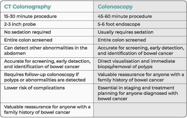
CT Colonography is a safe, effective, and considerably more comfortable alternative to traditional colonoscopy for bowel cancer screening - with no sedation required, a quicker procedure, and a faster recovery.
In New Zealand, access to conventional colonoscopy can be limited, meaning wait times are often long. CT Colonography offers a timely and reliable alternative, helping more women access bowel screening without unnecessary delay.
For women at low risk, CT Colonography (or colonoscopy) every five years is a sensible way to screen for bowel cancer and other changes to the colon and rectum. If you are at higher risk - due to family history, previous polyps, or other factors - it's worth discussing with your doctor whether to start earlier or screen more frequently.
Bowel cancer is highly treatable when caught early, which is exactly what regular screening is designed to do.
CT Colonography is a fast, accurate, and less invasive alternative to colonoscopy. See the comparison below for more information.
Low-dose CT (LDCT) is a quick, non-invasive scan that can detect lung cancer at an early stage - before symptoms develop, and when treatment options are at their broadest.
This is particularly relevant if you have a smoking history, whether you currently smoke or have quit in the past. Lung cancer often develops without early warning signs in people at higher risk, which is why proactive screening can make such a difference to outcomes.
The scan uses less radiation than a standard CT, requires no injections or sedation, and you can carry on with your day straight afterwards.
For anyone with a smoking history, LDCT offers a proactive and straightforward way to stay on top of lung health - providing reassurance when results are normal, and enabling earlier action when they're not. It works best as part of a broader, risk-based approach to your care, alongside medical advice from your doctor.
You may have heard that WB-MR is able to ‘check for everything’ and find disease early. While it can be used for screening or early detection in some situations it is not a complete test for all diseases, and it's important to understand its limitations.
Currently there is no evidence to demonstrate significant improvement to your health outcomes using whole‑body MRI as a screening tool. It can also identify incidental or low‑risk findings that may lead to unnecessary anxiety and follow‑up tests. In addition to this, not all diseases are detectable on whole‑body MRI - particularly in the early stages - which can offer you false reassurance.
Whole-body MRI may be appropriate:
- As part of specialist-led care or surveillance
- In high-risk genetic conditions such as Li-Fraumeni Syndrome (inherited TP53 loss)
- For specific medical reasons, such as inflammatory or systemic disease
We recommend screening that is targeted, and evidence based, matched to your own risk profile, and using imaging that’s known to improve outcomes (such as bowel, lung and the other screening options defined above).
If you believe a whole-body MRI may be appropriate for you, please discuss the potential benefits, limitations, and suitability of the test with your doctor and arrange a referral to book your appointment with us.
For women in their 50s and 60s, screening recommendations vary based on your personal risk profile. It's important to discuss this with your healthcare provider and have your risk assessed. From there you can consider the options that best suit your profile and arrange a referral.
Tomosynthesis
Tomosynthesis (also known as 3D mammography) captures multiple images from different angles, allowing the breast to be viewed in thin layers rather than a single flat image. This reduces the overlap of normal tissue, making it easier to identify small or subtle abnormalities that might otherwise be hidden - offering greater confidence in the results over standard mammography.
For women with an average risk profile, tomosynthesis every two years is recommended. For those with denser breasts (BIRADS C or D) or a moderate to high risk profile, annual tomosynthesis is recommended.
Contrast Enhanced Mammography
Contrast Enhanced Mammography (CEM) is a specialised mammogram that uses a small amount of contrast dye to highlight areas of increased blood supply in the breast. This can help detect cancers more clearly - particularly in dense breast tissue - by showing changes that may not be visible on standard mammography or tomosynthesis alone.
For women with denser breasts (BIRADS C or D) or a moderate to high risk profile, CEM is recommended every second year, alternating with tomosynthesis. This provides an additional layer of sensitivity and improves the chance of detecting cancers earlier.
MRI Breast
For women with denser breasts (BIRADS C or D) or a moderate to high risk profile, MRI may also be recommended periodically alongside annual tomosynthesis - or as an alternative to CEM. MRI uses a different technology, without radiation, that is more sensitive than mammography, making it better placed to pick up small or early abnormalities that may be difficult to see even with tomosynthesis.